










































i600 Scanning: Digital Rehabilitation
Clinical and laboratory routines demand accuracy. How many times does a job return with minor inaccuracies that consume time and resources? The uncertainty regarding the perfect fit of a mockup, the ideal adaptation of an occlusal splint, or the precision of an interocclusal record are silent pains that every dental professional has experienced. Each rework means a hidden cost, a delay in the schedule, and sometimes, a questioning of the process's predictability. It is in this scenario that the search for methods that guarantee greater control and predictability becomes not only relevant but essential.
📊 Quick Technical Summary
The MEDIT i600 Intraoral Scanner revolutionizes digital rehabilitation with its ability to record vertical dimension and intermaxillary position with millimeter precision. Integrated with software like Exocad, it allows for the planning and fabrication of mockups and printed splints with effective occlusal contacts, validating occlusion with comparative analysis of carbon points and digital scanning.
- Technology: Continuous 3D video (3D-in-motion)
- Speed: Up to 35 frames per second (FPS)
- Accuracy: Vertical dimension and intermaxillary registration
- Main Application: Prosthetic rehabilitation, occlusal planning, and splint fabrication.
The Reality of the Digital Workflow
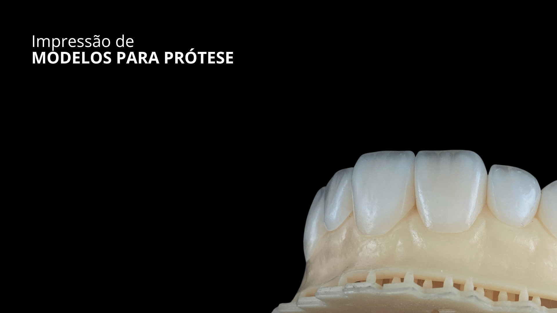
The perception that every step of treatment needs to be validated by concrete data is not new. What changes is the tool that allows this validation. A young patient undergoing complex rehabilitation needs solutions that offer longevity and perfect adaptation. Working with digital mockups and printed splints is already a reality, but the real difference lies in how these elements connect with the patient's mouth, and how occlusion is carefully adjusted. It's not enough to just scan; the scan must be a solid foundation for planning and execution.
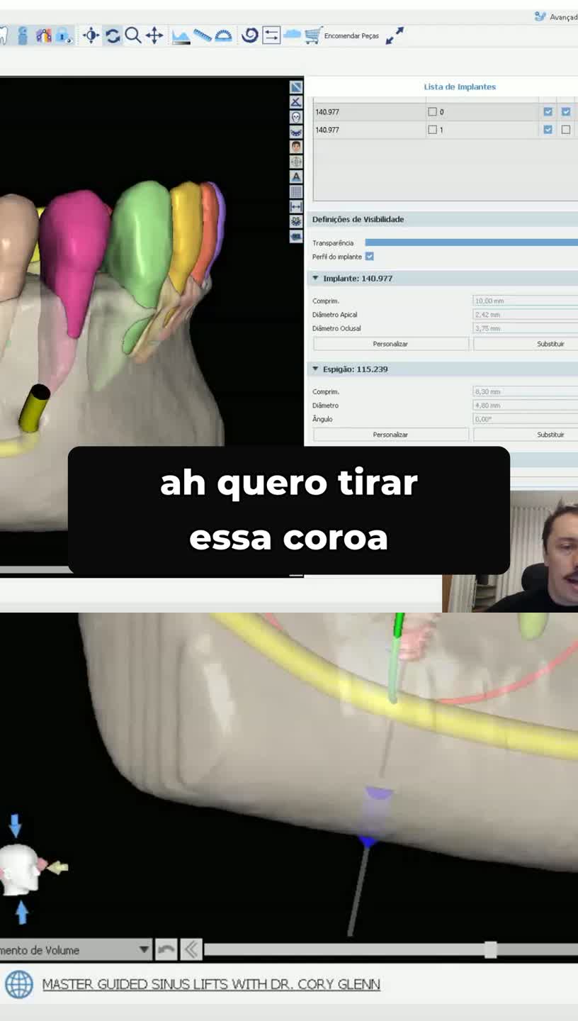
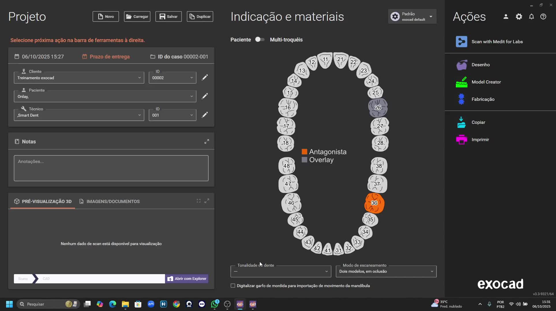
Precision in measuring vertical dimension is a critical point. Without it, all subsequent work can be compromised. The ability to accurately record the interarch distance, even in cases of vertical dimension opening that require auxiliary devices like the Gig, is a game-changer. This information, once captured, feeds into planning software like Exocad, allowing for detailed digital wax-ups and the fabrication of prosthetic pieces with the confidence that they were designed on a real and precise basis.
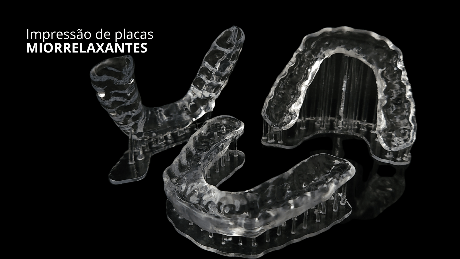
Leaving a finished case without adequate occlusal protection would be an unnecessary risk. The fabrication of a protective or stabilization splint, printed with effective contacts and adjusted on the same day the case is finalized, is not a luxury but a necessity. This agility and the quality of the printed piece, with a perfect fit and precise contact points, demonstrate the maturity of the digital workflow and the control the professional has over the clinical scenario.
Detailed Occlusal Analysis: From Carbon to Digital
Occlusal analysis has always been a challenge. The gomphosis, with its variation of 7 to 12 micrometers in isotonicity (light contacts), and 30 to 40 micrometers in isometry (clenching), shows how subtle and complex the interaction between teeth is. Understanding these nuances is crucial for precise adjustment. Carbon papers of different thicknesses, such as BK01 (200 micrometers) and BK28 (12 micrometers), or the metallic shimstock foil, are essential tools for identifying areas of contact and pressure.
The true innovation emerges when these physical tools are integrated into the digital workflow. By associating carbon marking in the mouth with scanning, it is possible to compare and verify the coincidence of pressure points. Using a thicker carbon, like BK01, to identify high-pressure areas, and then a thinner one, like BK28, to refine these contacts, and then scanning, allows for a visual and digital analysis of occlusion. The blue carbon points, superimposed on the red scan points, reveal areas of overcontour and high pressure, validating occlusal adjustment with an unimaginable level of detail.
This cross-validation between physical and digital provides a reliability map. The MEDIT i600 Intraoral Scanner, for example, offers a visualization that highlights areas where the scan is most accurate (in green), and points out those that need rescanning (in red). This functionality not only ensures mesh quality but also optimizes time, allowing the professional to focus only on areas that need correction, without redoing the entire process. It's clarity, security in investment, and predictable financial return.
The ability to adjust precision parameters, such as 40 micrometers for isometry (clenching) and 7 to 12 micrometers for isotonicity (light contact), directly in the software, allows for a meticulous verification of occlusion. The coincidence between physical carbon points and virtual points in the scan, validated by studies and clinical practice, offers a level of detail that transforms occlusal analysis. It is not just a diagnostic tool, but a complete control and verification system.
MEDIT i600 Intraoral Scanner: The Heart of the Digital Workflow
MEDIT i600 Intraoral Scanner
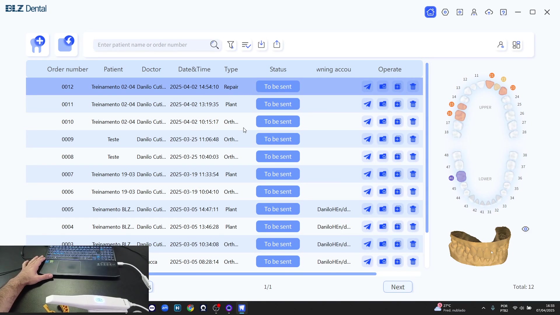
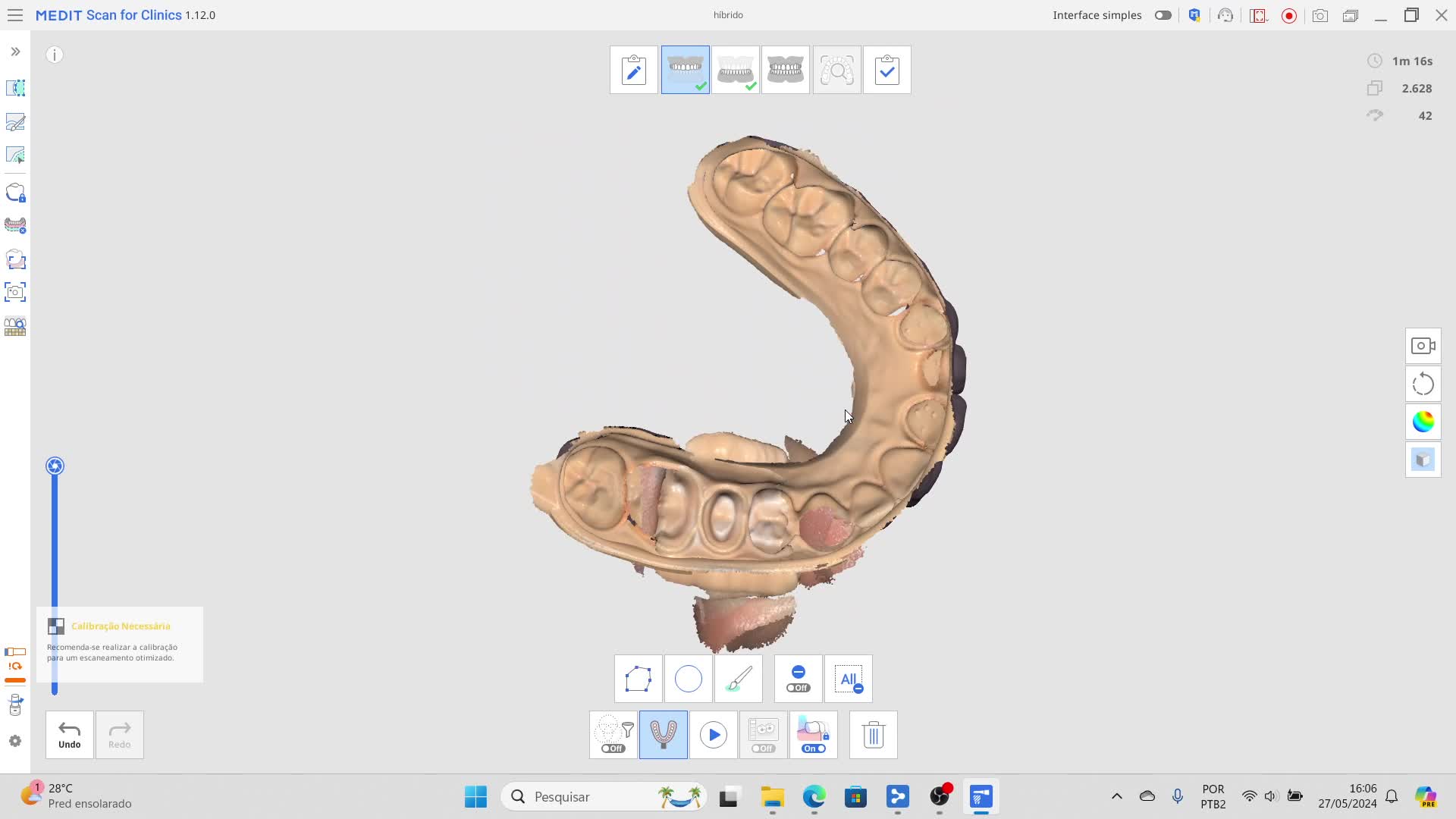
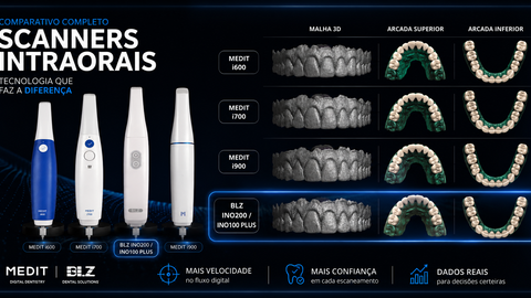
The MEDIT i600 Intraoral Scanner is the tool that consolidates the transition to digital dentistry. With its continuous 3D video technology (3D-in-motion), it captures up to 35 frames per second (FPS) in vibrant 8-bit colors, ensuring superior anatomical reading. Its fully open architecture is a crucial differentiator, allowing the export of files in STL, OBJ, and PLY formats. This ensures compatibility with a wide range of CAD/CAM software and dental 3D printers on the market, eliminating ties and additional costs. Weighing only 245g with the tip and featuring an ergonomic design, the i600 minimizes operator fatigue and provides greater patient comfort. The 180° reversible tip facilitates access to all areas of the mouth, optimizing the scanning experience. In addition, the tip is autoclavable for up to 100 cycles, reinforcing biosafety practices. Free of monthly fees or recurring software costs, the MEDIT i600 Intraoral Scanner is a smart investment to modernize dental practice. For more details on how this scanner can transform your clinic, consult the purchase URL.
- Technology: Continuous 3D video (3D-in-motion)
- Speed: Up to 35 frames per second (FPS)
- Colors: 8-bit
- Weight: 245g (with tip)
- Tip: 180° reversible, autoclavable (up to 100 cycles)
- Export Formats: STL, OBJ, PLY (open architecture)
- Cost: Free of monthly fees or recurring software costs
Oclusense and Occlusal Analyzer System: Dynamic Analysis in Practice
Associating the precision of the Medit i600 with an occlusal analysis system like Oclusense elevates the level of practice. Oclusense offers pressure analysis at 256 points, with a ranking that allows identification of occlusal imbalances. This ability to evaluate pressure in detail, in real time, transforms how the professional approaches occlusal adjustment. The evolution is clear: from physical carbon to digital, with the possibility of seeing the exact pressure at each contact point.
The functionality of Occlusal Analyzer, free software for Medit users, even allows dynamic analysis within the scanner itself. This means that no additional equipment is needed to obtain crucial information about occlusion in motion. The ability to perform isotonic and isometric tests, observing the variation of pressure and the coincidence of contact points between physical and virtual, offers a complete and in-depth view of the patient's occlusal relationship.
The combination of high-precision scanning, digital planning, and advanced occlusal analysis tools allows the professional to control the entire process, from diagnosis to case completion. This translates into greater safety for the patient and superior clinical results. Digital dentistry is no longer a promise, but a reality that offers control, predictability, and excellence in daily clinical practice.
✅ Control and Predictability in Digital Dentistry
The decision to incorporate digital dentistry into practice is not just about technology, but about the pursuit of more consistent results and a more efficient workflow. The ability to accurately record vertical dimension, plan mockups and splints in software, and validate occlusion with the integration of carbon and scanning, offers a level of control that was previously unattainable. It is the certainty that each step of the treatment is being executed with maximum precision, minimizing surprises and optimizing time. Choosing a system that offers this integration and reliability is a conscious and strategic decision for the professional seeking excellence and safety in their practice.

About the author
Prof. Dr. Weber Adad Ricci
Reabilitação Oral · Prótese Dentária · Odontologia Digital · UNESP FOAr
Doutor em Reabilitação Oral/Prótese pela FOAr/UNESP (2005). Professor RTC MS-3.1 — Depto. Odontologia Social, UNESP. Autor dos livros Lógica 01 e 02 (11.000+ cópias, 40+ países, 3 idiomas). Criador da Matriz BRB e do conceito Estética e Oclusão Bioinspiradas. Fundador do Instituto Weber Ricci (3.000+ alunos). Consultor Smart Dent · Medit · Ultradent · GC · Kavo/Kerr. ORCID: 0000-0003-0996-3201 · Scopus: 55509352400.