














3D and Color Alveolar: Agility in Diagnosis
📊 Quick Technical Summary
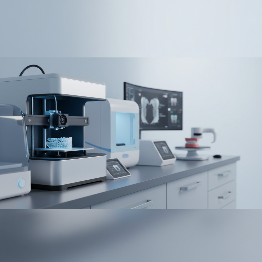
Colored 3D visualization of alveoli optimizes diagnosis with precision and agility, allowing for detailed analysis of bone structure and soft tissues.
- Technology: Colored 3D printing
- Benefit: Precise and fast diagnosis
- Application: Alveolar analysis and extraction planning
- Advantage: Optimization of workflow and patient communication
Opening – Obvious Truths
In the daily routine of a clinic or laboratory, time is a resource that is never abundant. The pressure for accurate diagnoses and effective treatment plans, often with tight deadlines, is constant. How many times has the lack of clear and immediate visualization delayed a crucial decision or generated insecurity in planning?
The need to have all information visible, in an understandable and quick way, is a silent pain that affects efficiency and, at times, the trust the patient places in the professional. Rework, caused by interpretation or communication failures, is a hidden cost that accumulates.
Development – Progressive Agreement
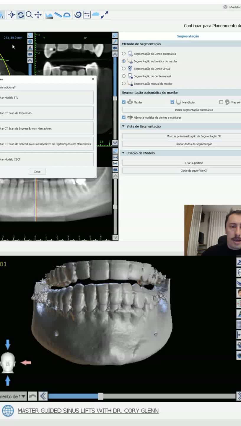
It is a reality that the complexity of certain structures, such as the alveolar region, requires a level of detail that two-dimensional images cannot always deliver. The interpretation of radiographs and tomographies is fundamental, but the mental transposition to a three-dimensional model can be challenging and consume valuable time.
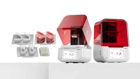
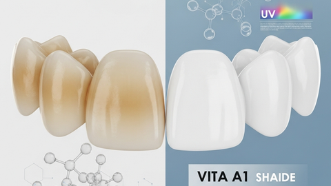
Think about the difference it makes to literally have in your hands a model that faithfully reproduces the patient's anatomy, with the differentiation of soft and hard tissues by color. This eliminates subjectivity and the need for multiple verifications, generating an instant “this is true” for the person analyzing it.
The ability to manipulate and explore a physical, colored, three-dimensional model allows for clear identification of the position of nervous structures, bone thickness, and the relationship with adjacent teeth. This level of detail is visible, tangible, and undeniably useful.
Natural Insertion of the Solution
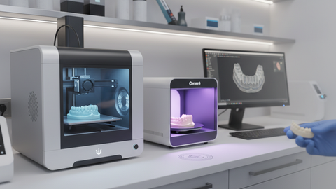
In this context, the possibility of generating a 3D model with colors, representing the alveolar ready in a matter of seconds, emerges as a natural evolution. It is not an empty promise, but a tool that fits perfectly with the need to optimize workflow and improve diagnosis.
Digital dentistry, by allowing 3D printing of anatomical models with such richness of detail, transforms the way professionals face daily challenges. It is technology at the service of clarity and efficiency, without circumlocution or unnecessary complications.
Integration of Testimony
For the professional who experiences this routine, the ability to have an alveolar model ready in seconds, and with color differentiation, means a revolution. It means, for example, that the planning of a complex extraction can be done with much more safety and predictability.
Colored visualization allows for more effective communication with the patient, who then better understands the procedure and its steps. This clarity in explanation strengthens the relationship of trust and facilitates treatment acceptance, transforming a previously abstract process into something concrete and understandable for everyone.
The decision to incorporate this technology is not a gamble, but a logical step for those seeking more agility and precision, aligning with the evolution of clinical practice.
Clarity and Decision Block
It's clarity, it's investment security and predictable financial return.
Closing – Silent Authority
The choice of tools that add real value to diagnosis and planning is a reflection of professional maturity. Opting for solutions that bring more visibility and precision, without artifice, is the most solid way to build a practice of excellence and recognition. The decision is conscious, informed, and focused on the best delivery to the patient.

![[COMPARATIVO] - Impressora 3D qual a ideal para chairside print](https://b-vz-004839ee-19a.tv.pandavideo.com.br/8589e7fc-982f-4a12-a303-3f96a31fbee8/thumbnail.jpg)